The accuracy of the automated image analysis depends on image quality. It is thus a smart time investment to optimize sample preparation and image capturing!
As with any image analysis tool the quality of the produced data depends much on the quality of the analyzed images. With a poor-quality anatomical sample and image the manual editing may easily require 10x longer and more, while the achieved data quality may still be insufficient. Details on the proper sample preparation and digitizing can be obtained in the references below. In short, the following points should be considered:
a) Collecting samples in the field
- Use sharp tools to avoid cracks
- Proper orientation of samples (usually perpendicular to the axial direction of xylem cells)
b) Preparing anatomical sections *
- Use sharp microtome blades
- Cut with proper orientation of samples (usually perpendicular to the axial direction of xylem cells)
- Thick sections can result in systematic over- or underestimation of measured anatomical features; in any case, always use the same thickness.
c) Digitizing anatomical samples *
- Use a sufficient magnification (conifers: typically 10x objective, angiosperms: typically 4x objective)
- Adjust microscope settings (illumination, aperture, focus); keep the same settings for all samples
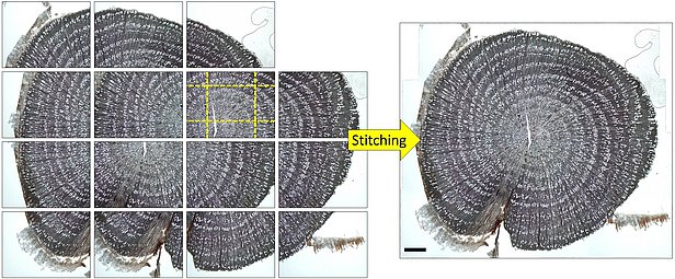
- Use specialized stitching software such as PTGui (www.ptgui.com) to create high-resolution images of entire anatomical samples (see Fig. 1 below); a PDF protocol for stitching with PTGui can be obtained here.
- Note: slide scanners can produce high-quality and high-resolution images of entire anatomical samples
* Relatively large anatomical features such as earlywood vessels in ring-porous species can be analyzed in images captured with a flatbed scanner at 1500-2500 dpi directly from the prepared wood surface.
References 
- von Arx G, Crivellaro A, Prendin AL, Cufar K, Carrer M. 2016. Quantitative wood anatomy - practical guidelines. Frontiers in Plant Sciences 7:781. http://journal.frontiersin.org/article/10.3389/fpls.2016.00781
- Gärtner H, Cherubini P, Fonti P, von Arx G, Schneider L, Nievergelt D, Verstege A, Bast A, Schweingruber FH, Büntgen U. 2015. Technical challenges in tree-ring research including wood anatomy and dendroecology. Journal of Visualized Experiments. e52337, doi:10.3791/52337. http://www.jove.com/video/52337
- von Arx G, Stritih A, Čufar K, Crivellaro A, Carrer M. 2015. Quantitative Wood Anatomy: From Sample to Data for Environmental Research. doi: 10.13140/RG.2.1.3323.0169. URL: https://www.youtube.com/watch?v=gz_mJVmn-vQ&feature=youtu.be
Fig.1: Illustration how PTGui can be used to stitch overlapping images to a single high-resolution image of the entire anatomical sample (see PDF protocol). The overlap with the neighboring images is visualized for one of the images with yellow dashed lines. Verbascum thapsus root cross-section. Scale bar: 1 mm. Adjusted from von Arx et al. 2016, FPS
