On this page you can find a collection of ROXAS configuration files. Use a configuration file whose thumbnail image resembles your own samples as a starting point for customizing your own configuration.
Instruction 
- Download one to several configuration files (*.rsf) and copy-paste them into the configuration folder (in ROXAS main window, push 'Manage configuration files' to access the folder)
- Re-launch ROXAS. The added configuration(s) will appear in the configuration list in the ROXAS main window.
- Adjust the configuration file(s) of choice as described in the ROXAS Manual.
Note: Configurations are rather specific! They are usually optimized for all of the following: the anatomy of a given species, the plant organ, the sample processing and staining technique, the image capturing system (microscope, camera, scanner), and the image resolution.
Using configuration files with ROXAS 
Any new version of ROXAS is able to use an old configuration file, however don't use a new configuration file with an older ROXAS version! As ROXAS is developing, new settings are added to the configuration files, which an older ROXAS version does not support.
Contribute yourself 
If you defined a new configuration that works well on your samples and you want to see it on this page, please contact me!
Download all configuration files
>>> Download all configuration files (last updated: 1 April 2016) <<<
Individual configurations 
Angiosperms 
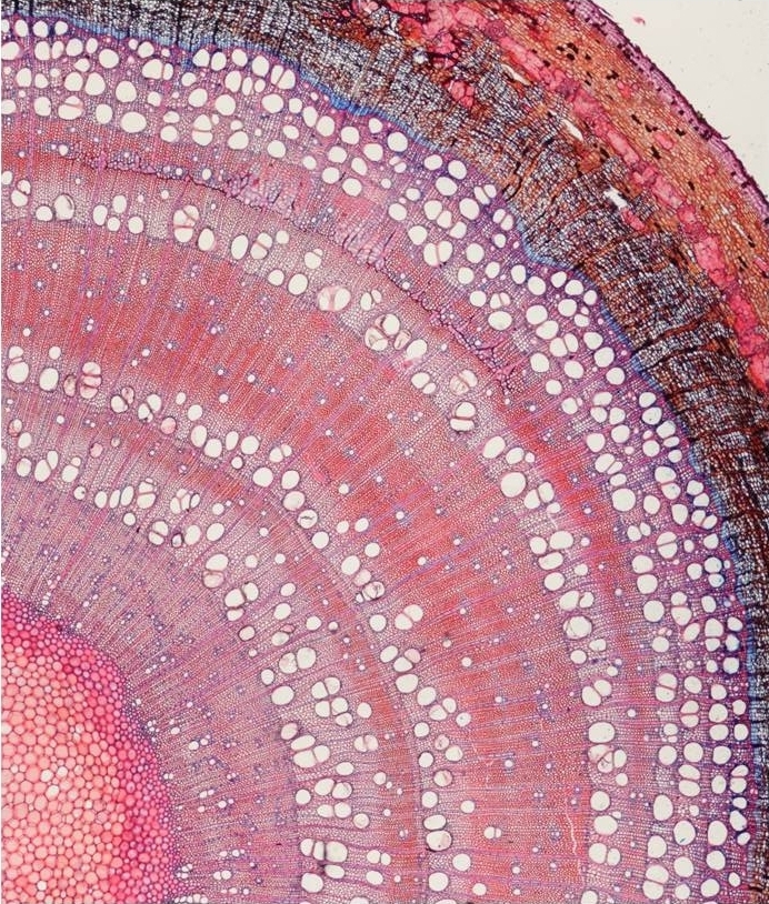
Configuration: Fraxinus_excelsior_0.945.rsf
Species: Fraxinus excelsior
Date: 4 January 2017
Tissue: Branch
Tissue age: Young
Type: Circular
Sample processing: Thin sections
Staining: Safranin – astra blue
Image capturing: Transmitted-light microscope
Image resolution: 0.945 pix / µm
Conduit area 150-15,000 µm2
Ring detection configured: No
Image credit: Georg von Arx

Configuration: Acacia_greggii_0.752.rsf
Species: Acacia greggii
Date: 1 April 2016
Tissue: Root collar
Tissue age: Seedling
Type: Circular
Sample processing: Thin sections
Staining: Phloroglucinol-HCl
Image capturing: Transmitted-light microscope
Image resolution: 0.752 pix / µm
Conduit area: 500-4500 µm2
Ring detection configured: No
Image credit: Steven Woods
Betula pubescens 

Configuration: Betula_pubescens_0.988.rsf
Species: Betula pubescens
Date: 1 April 2016
Tissue: Stem
Tissue age: Mature
Type: Linear
Sample processing: Thin sections
Staining: Methylene blue
Image capturing: Transmitted-light microscope
Image resolution: 0.988 pix / µm
Conduit area: 170-4000 µm2
Ring detection configured: No
Image credit: Marina Bryukhanova
Configuration: Betula_nana_0.945.rsf
Species: Betula nana
Date: 1 April 2016
Tissue: Stem
Tissue age: Mature
Type: Circular
Sample processing: Thin sections
Staining: Safranin – astra blue
Image capturing: Transmitted-light microscope
Image resolution: 0.945 pix / µm
Conduit area: 500-8000 µm2
Ring detection configured: No
Image credit: Sigrid Nielsen

Configuration: Fagus_sylvatica_stem_0.456.rsf
Species: Fagus sylvatica
Date: 1 April 2016
Tissue: Stem
Tissue age: Young tree
Type: Linear
Sample processing: Thin sections
Staining: Safranin – astra blue
Image capturing: Transmitted-light microscope
Image resolution: 0.456 pix / µm
Conduit area: 40-6000 µm2
Ring detection configured: No
Image credit: Britta Eilmann
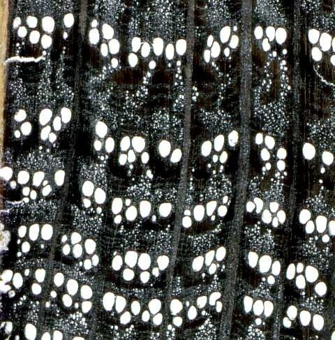
Configuration: Quercus_robur_1500dpi.rsf
Species: Quercus petraea & robur
Date: 1 April 2016
Tissue: Stem
Tissue age: Mature
Type: Linear
Sample processing: Surface preparation
Staining: Black marker, chalk powder
Image capturing: Scanner
Image resolution: 0.059 pix / µm (1500 dpi)
Conduit area: 1000-140,000 µm2
Ring detection configured: Yes
Image credit: Patrick Fonti

Configuration: Salix_polaris_0.752.rsf
Species: Salix polaris
Date: 1 April 2016
Tissue: Stem
Tissue age: Mature
Type: Circular
Sample processing: Thin sections
Staining: Safranin – Astra blue
Image capturing: Transmitted-light microscope
Image resolution: 0.752 pix / µm
Conduit area: 400-8000 µm2
Ring detection configured: No
Image credit: Agata Buchwal
Conifers 

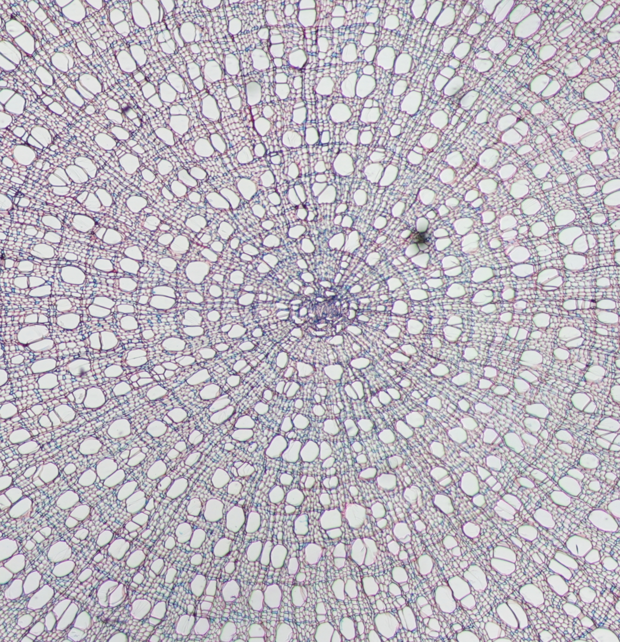
Configuration: Larix_decidua_2.074.rsf
Species: Larix decidua
Date: 1 April 2016
Tissue: Branch
Tissue age: Small treeline tree
Type: Linear
Sample processing: Thin sections
Staining: Safranin
Image capturing: Transmitted-light microscope (100x)
Image resolution: 2.074 pix / µm2
Conduit area: 5-1000 μm2
Ring detection configured: Yes
Image credit: Angela Luisa Prendin

Configuration: Picea_abies_0.833.rsf
Species: Picea abies
Date: 1 April 2016
Tissue: Stem
Tissue age: Mature
Type: Linear
Sample processing: Thin sections
Staining: Safranin
Image capturing: Transmitted-light microscope (40x)
Image resolution: 0.833 pix / μm
Conduit area: 5-2000 μm2
Ring detection configured: Yes
Image credit: Giai Petit

Configuration: Pinus_leucodermis_0.833.rsf
Species: Pinus leucodermis
Date: 1 April 2016
Tissue: Stem
Tissue age: Mature
Type: Linear
Sample processing: Thin sections
Staining: Safranin
Image capturing: Transmitted-light microscope (40x)
Image resolution: 0.833 pix / μm
Conduit area: Yes
Ring detection configured:
Image credit: Marco Carrer

Configuration: Pinus_sylvestris_2.083.rsf
Species: Pinus sylvestris
Date: 1 April 2016
Tissue: Stem
Tissue age: Mature
Type: Linear
Sample processing: Thin sections
Staining: Safranin
Image capturing: Transmitted-light microscope (100x)
Image resolution: 2.0830 pix / μm
Conduit area: 5-2000 μm2
Ring detection configured: Yes
Image credit: Marco Carrer

Configuration: Pinus_sylvestris_2.3613.rsf
Species: Pinus sylvestris
Date: 1 April 2016
Tissue: Stem
Tissue age: Mature
Type: Linear
Sample processing: Thin sections
Staining: Safranin-Astrablue
Image capturing: Transmitted-light microscope (100x)
Image resolution: 2.3613 pix / μm
Conduit area: 5-2000 μm2
Ring detection configured: Yes
Image credit: Georg von Arx
Needles 

Configuration: Needles.rsf
Species: Pinus sylvestris
Date: 1 April 2016
Tissue: Needles
Tissue age: Branch of mature tree
Type: Linear
Image capturing: Flatbed scanner
Settings: Transparency
Image resolution: 300 dpi (11.811 pix / mm)
Needle area: 2-500 mm2
Image credit: Georg von Arx