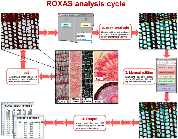
ROXAS is designed to allow efficient and flexible analysis of anatomical features in the xylem and other tissues including phloem, rays and also conifer needles. The following features are included (see overview in Fig. 1).
a) Automatic image processing
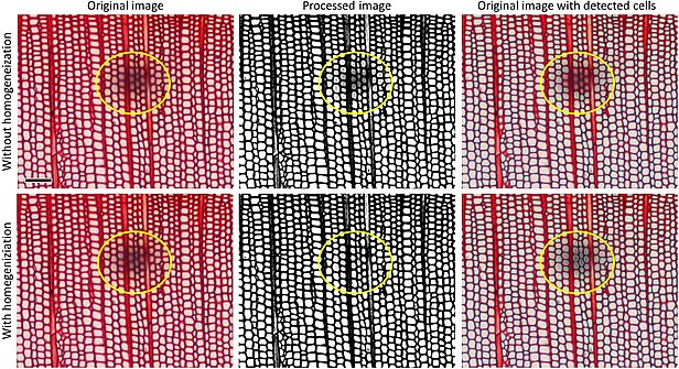
- Image improvement for inhomogeneous and poor contrast (see Fig. 2 below)
- Edge enhancement
b) Automatic cell and ring detection
- Automatic recognition of annual rings and cells in large samples (linear and circular) with >100 annual rings and 1,000,000 cells
- Cell detection and filtering based on color, size, shape & context
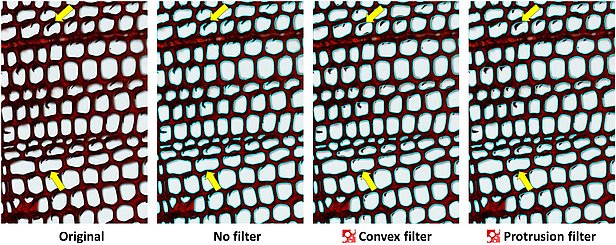
- Automatic filters to remove particles within cells and protrusions from cell walls (see Fig. 3 below)
c) Efficient manual editing of cells and ring borders
- All measurements visualized as editable vector overlays
- Selecting areas of interest (AOI) / exclusion (AOE)
- Toolbox for automatic cell filtering using complex criteria
- Deleting, drawing, correcting, undoing tools of cell lumen outlines and ring borders
- Quality controlling sorted list of largest and smallest cells
d) Data output
- Exhaustive set of output parameters
- Data output in well-organized MS Excel (see example file, 2.9 MB) and text files
- Summarizing function to copy and paste data from all analyzed images into one file
e) Batch processing
- Batch processing functions for all relevant steps to avoid waiting time for the user (spatial calibration, project preparation and execution, etc.)
f) Customizing analysis
- Large collection of pre-defined configurations for many samples (ROXAS Configuration Library)
- Adjusting and creating own configurations to specifics of samples
- Customizing colors of vector overlays (cells, ring borders, AOE, AOI)
g) Additional features
- Analysis of length (considering curvatures!), width and area of scanned conifer needles
- Analysis of tissue such as rays (currently only in circular cross-sections)
- User manual (115 p.)


